
Cardiac Arrhythmias in Dogs and Cats

How does the heart know when to beat?
In order to pump blood to the lungs and body, the heart must work in a step-by-step coordinated fashion. The heart has an electrical conduction system that is responsible for controlling the heart rate and rhythm. This electrical conduction system generates electrical impulses (waves), which travel through the heart, stimulating the heart’s muscles to contract and push blood through the arteries and out into the body.
Step 1: Impulse generation in the SA node
The sinus node, or sinoatrial (SA) node, is a clustered collection of similar cells located in the top of the right atrium (the first chamber that blood enters as it returns from traveling through the rest of the body), its purpose being to generate electrical impulses and to serve as the heart’s pacemaker.
Step 2: SA node to AV node impulse conduction
The other node in the heart is called the atrioventricular (AV) node. Like the SA node, the AV node is a clustered collection of similar cells situated in the bottom of the right atrium, close to the ventricle (the chamber that receives blood from the atrium). The AV node receives impulses from the SA node.
Step 3: AV node to ventricles impulse conduction
After a small delay in the AV node, the AV node then directs the impulses to the ventricles. This delay allows for the atrium to eject blood into the ventricle before the ventricular muscles contract. The AV node can also take the place of the SA node as the heart’s pacemaker if the SA node should malfunction because of disease.
What is a cardiac arrhythmia?
If you were to listen to your heart with a stethoscope, the “lub-dub” (heart beat) you hear is the sound of the heart valves closing. This corresponds to the ventricular muscle contracting. The “lub” consists of the left and right atrioventicular valves closing. The left cardiac valve is the mitral valve and the right cardiac valve is the tricuspid valve. The “dub” consists of the closure of the pulmonary and aortic valves after the ventricles have ejected blood into the pulmonary artery (large vessel supplying the lungs) and aorta (large vessel supplying the body), respectively.
Arrhythmias are abnormal heart rhythms. Some arrhythmias are normal variations (such as the respiratory sinus arrhythmia in dogs). Dangerous arrhythmias are those that result in clinical signs and/or put the animal at risk for sudden cardiac death.
What are types of arrhythmias?
Sinus node disease results in too low of a heart rate (bradycardia). One of the diseases affecting the sinus node is sick sinus syndrome (SSS). Older miniature Schnauzers, Cocker Spaniels, and West Highland White Terriers are the most commonly affected breeds.
Atrial standstill is a condition where normal atrial tissue has been replaced with abnormal tissue and cannot conduct the electrical impulse properly. This results in bradycardia. English Springer Spaniel is the breed most commonly affected by this disease.
Heart block (3rd degree AV block) is a type of arrhythmia that results in bradycardia and is due to disease of the AV node (the top chambers of the heart cannot communicate with the lower chambers of the heart). Commonly affected breeds include Chow Chow, Cocker Spaniel, German Wirehaired Pointer, Pug and Labrador Retriever.
Ventricular premature complex (VPC, aka ventricular premature contraction) is a type of irregular heartbeat. An electrical impulse is initiated within the ventricles instead of the SA node, causing the ventricles to contract too early (thus the premature in ventricular premature complex). Ventricular tachycardia exhibits a rapid heart rate due to many VPCs firing rapidly, often resulting in symptoms such as exercise intolerance or collapse.
Supraventricular premature complexes arise either from the tissue in the atrium or the tissue surrounding and in the AV node. Supraventricular tachycardia also exhibits a rapid heart rate due to supraventricular premature complexes firing rapidly.
Atrial fibrillation (Afib) is the most common supraventricular tachycardia in dogs and is often the result of an underlying cardiac condition. It is a fast, irregular heart rhythm that, if heard with a stethoscope, sounds like “tennis shoes in a dryer”. Irish Wolfhound is a breed that is predisposed to “lone Afib” in which the arrhythmia is present in the absence of underlying heart muscle disease.
What are causes of arrhythmias?
Cardiac causes of arrhythmias include: Heart muscle disease (such as dilated cardiomyopathy, hypertrophic cardiomyopathy and arrhythmogenic right ventricular cardiomyopathy), congenital heart defects (especially subaortic stenosis), severe valve leakage and enlargement of the cardiac chambers (chronic degenerative mitral valve disease), myocarditis (inflammation of the heart muscle), trauma to the heart muscle (such as an animal being hit by a car), age-related changes, and replacement of the heart muscle with inflammatory cells or cancer cells.
Non-cardiac causes of arrhythmias include: Gastric dilation and volvulus (stomach turns and flips on itself), inflammation of the pancreas, low blood magnesium, severe anemia (low red blood cell count); diseases of the spleen, liver or GI tract; neurologic disease (i.e. brain tumors); endocrine disease (i.e., of the thyroid gland, adrenal glands); muscular dystrophy, anesthetic agents, medications, and toxins (i.e., chocolate intoxication).
What are the symptoms of arrhythmias?
- Weakness
- Collapse/fainting
- Exercise intolerance
- Fluid accumulation in the abdomen (bloating)
- Increased respiratory rate
- Labored breathing
- Sudden death
However, it is not uncommon for dogs and cats to appear outwardly normal (no symptoms) despite having a cardiac arrhythmia.
How are arrhythmias diagnosed?
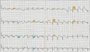
Your family veterinarian will diagnose an arrhythmia when listening to the heart with a stethoscope or on an EKG reading. An EKG (electrocardiogram or ECG) is the best diagnostic test to identify and diagnose the specific type of arrhythmia. A Holter monitor is an ambulatory EKG (the dog can wear it home and the device records the cardiac rhythm over the next 24-48 hours) that determines the frequency (how often the arrhythmia occurs) and severity of the arrhythmia. Your family veterinarian will then recommend treatment and/or referral to the Veterinary Cardiologist, if the arrhythmia appears dangerous on the EKG or the Holter monitor.
How are arrhythmias treated?
A pacemaker (a surgical procedure performed by a Veterinary Cardiologist) is the standard recommended treatment for sick sinus syndrome, atrial standstill and complete heart block.
Various antiarrhythmic drugs are utilized for supraventricular and ventricular arrhythmias. Intravenous drugs are used for life threatening arrhythmias and oral drugs for long term therapy. Uncommonly, certain supraventricular arrhythmias can be treated with radiofrequency ablation, while others can be treated with electrocardioversion (i.e., lone Afib). Defibrillation is indicated immediately for life threatening ventricular arrhythmias (ventricular fibrillation).
What is the prognosis for a dog or cat with an arrhythmia?
The prognosis is highly variable depending on what type of arrhythmia is present and if there is a non-cardiac and treatable cause versus underlying severe heart disease (i.e., dilated cardiomyopathy in Doberman Pinschers). Pacemaker implantation for a slow heart rate (3rd degree AV block and sick sinus syndrome) is associated with a good prognosis in the absence of severe underlying cardiac disease.
Edited by:
Rebecca Saunders, DVM, DACVIM (Cardiology)
April, 2020
Articles by Specialty
- Cardiology (19)
- Large Animal Internal Medicine (23)
- Neurology (18)
- Oncology (22)
- Small Animal Internal Medicine (29)
Articles by Animal
- Cats (35)
- Dogs (52)
- Farm Animals (5)
- Horses (12)