
Pacemaker Therapy

What is a pacemaker?
Pacemakers are small devices that are implanted in a dog or cat’s heart that can increase their heart rate. They consist of two parts: the generator and the lead wire. The pacemakers that are used in veterinary patients are to prevent the heart rate from getting too low, unlike some used in human patients that can slow the heart rate down as well. Pacemakers function by delivering small shocks to the heart muscle to stimulate the heart to beat. Modern pacemakers are small with most being about the size of a silver dollar and the thickness of a pencil.
When does a dog or cat need a pacemaker?
Pacemakers are needed when the heart rate becomes too slow and the patient either becomes very lethargic, faints, or both. Some patients can die if their heart rates gets too low. The heart rhythms that can cause these conditions are most frequently:
- Atrio-ventricular block (AV block)
- Sick Sinus Syndrome or Sinus Node Dysfunction (SSS/SND)
- Atrial standstill
Medical therapy can be tried to correct these rhythms, but it is rarely successful. Many patients are somewhat elderly when they need a pacemaker, yet pacemakers can be placed successfully in these patients as well. Bloodwork will be required to rule out some other medical issues that can cause slow heart rates.
What breeds are most likely to have abnormally slow heart rhythms that may require a pacemaker?
- West Highland White Terriers
- Miniature Schnauzers
- Cocker Spaniels
However, any breed of dog or cat can require a pacemaker.
How is the pacemaker surgery done?
In order to place a pacemaker in a pet:
- General anesthesia is required
- A temporary pacing unit is placed to make sure that the heart rate does not go too low during anesthesia.
- There are two methods of placing a permanent pacemaker
- Transvenous: A small incision is made in the neck and the pacemaker lead is guided down the jugular vein into the bottom of the right ventricle via video x-ray called fluoroscopy. The pacemaker generator is then placed under the muscles of the neck.
- Epicardial: This is usually done in cats and some very small dogs. The lead is attached to the outside of the heart via an incision in the belly and the diaphragm. The pacemaker generator is then placed inside the abdomen/belly.
What are the potential complications of pacemaker placement?
Pacemaker surgery is generally successful but significant complications can occur in up to 33% of cases. The most common complications include:
- Lead dislodgement (the pacemaker lead fails to remain in contact with the heart muscle and does not generate an appropriate heartbeat)
- Infection
- Lead fracture (the pacemaker lead develops a short circuit in its wiring)
- End of battery life. Most pacemaker batteries last 5-10 years depending on how much the patient requires the pacemaker to help its heartbeat. Batteries/generators can be easily replaced with minor surgery.
What follow-up is required after pacemaker placement?
- A recheck with your cardiologist is usually recommended 2-4 weeks after placement of the pacemaker. During this time period, it will be very important to keep your pet calm, so they do not dislodge the lead wire from inside the heart. After about 4 weeks, the pacemaker lead wire will be scarred down to the heart muscle.
- Twice yearly interrogation of the pacemaker generator to assure the device is functioning correctly for your pet
- Avoid strong magnets around the neck. Microwaves are okay.
- Leash walking with a harness instead of a collar or choker collar is recommended to avoid putting pressure on the pacemaker that has been implanted in the neck region.
- Your cardiologist may also want to repeat an echocardiogram periodically especially if your pet had an enlarged heart prior to the pacemaker surgery
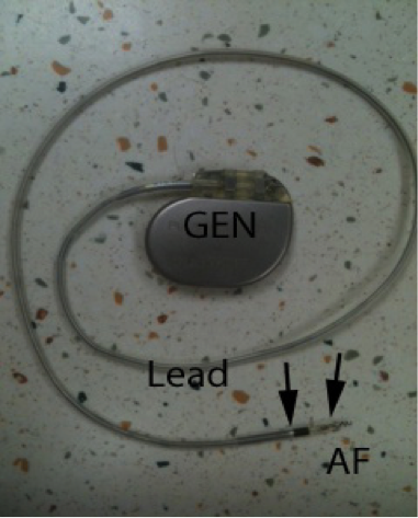
Figure 1. Pacemaker generator (GEN), lead and active fixation tip (AF) with bipolar electrodes (arrowheads).
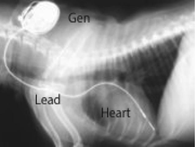
Figure 2. X-ray of a small dog showing the pacemaker generator in the neck (Gen) and the pacemaker lead (Lead) extending from the generator through the neck and into the heart. For orientation purposes, the spine runs along the top of the picture the breast bone runs along the bottom of the picture and the front legs are under the lead legend. The lead is the white structure running from the generator into the heart.
REFERENCES
Moise NS: Pacemaker Therapy. In Fox PR, Sisson D, Moise NS (eds): Textbook of Canine and Feline Cardiology. Philidelphia, Saunders, 1999, pp 400-425.
Bright JM. Pacemaker Therapy. In Smith FWK JR, Tilley LP, Oyama MP, Sleeper MM eds: Manual of Canine and Feline Cardiology, 5th ed. St Louis, MO, 2016.Elsevier, pp 382-393.
...
Edited by:
Bill Tyrrell, DVM, DACVIM (Cardiology)
April, 2020
Articles by Specialty
- Cardiology (19)
- Large Animal Internal Medicine (23)
- Neurology (18)
- Oncology (22)
- Small Animal Internal Medicine (29)
Articles by Animal
- Cats (35)
- Dogs (52)
- Farm Animals (5)
- Horses (12)