
Mast Cell Tumors in Dogs and Cats | Canine and Feline MCT

What is a mast cell tumor?
Mast cell tumors (MCT) represent a cancer of mast cells, which are a type of white blood cell involved in allergy and inflammation. Mast cell tumors are the most common skin tumor in dogs. Mast cells contain granules filled with substances that can be released into the bloodstream. In large amounts, these substances can cause systemic (body-wide) complications including stomach ulcers and bleeding and low blood pressure.
Older dogs are most commonly affected. Some predisposed breeds of dogs include Boxers, Boston terriers, Bulldogs, pugs, Labrador retrievers, golden retrievers, Weimaraners, Rhodesian Ridgebacks, cocker spaniels, schnauzers, Staffordshire Terriers, beagles and Shar Peis.
The cause of MCT remains unknown. Because certain breeds of dogs are predisposed to this cancer, genetics are thought to play a role. Additionally, there are known molecular abnormalities associated with MCT. Chronic inflammation may also play a role.
What are the signs and symptoms of mast cell tumors?
Mast cell tumors can vary in appearance.
- These tumors typically present as a lump on or under the skin.
- The lump can be hairless or covered by hair, and/or red, ulcerated or swollen.
- These lumps are often itchy.
- A history of waxing and waning size of the tumor can be reported. This is due to the fact that mast cells contain histamine in their granules and this can be released to cause swelling.
Some MCT can have a more benign-type of behavior where the tumor is present for months to years without change in size or a more aggressive behavior where growth occurs rapidly. If these tumors spread, the typical behavior is first involvement of “local” lymph nodes near the tumor followed by involvement of the liver or spleen.
How is a diagnosis of mast cell tumor made?
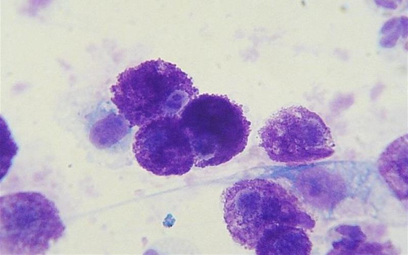
Initial evaluation of a skin mass tends to start with a fine needle aspirate (also called cytology). This is a non-invasive method to extract cells from a mass to obtain a diagnosis. Mast cell tumors have a very specific appearance that should make diagnosis straightforward with cytology.
Following diagnosis of MCT, additional diagnostics performed include:
- Blood work
- Fine needle aspirate of the local lymph node
- Abdominal ultrasound
- Chest radiographs (x-rays)
Photo: Cytology of a mast cell tumor from a Labrador Retriever. Image by Joel Mills.
How are mast cell tumors treated?
Surgical removal of the MCT is the treatment of choice. This allows for removal of the mass and also allows for information about prognosis (determining the grade of the tumor). Prior to surgery, patients are sometimes placed on antihistamines (such as Benadryl) and antacids (such as Pepcid AC or Prilosec) to alleviate side effects from the histamine that is present in the MCT granules.
The most important considerations when determining if a patient needs additional treatment following surgery are:
- Tumor grade
- Completeness of surgical removal (determined by evaluation of the tissue by a pathologist)
- Determination whether the MCT has spread or not
Following removal of the MCT, all tissue removed should be submitted to a pathologist at a diagnostic laboratory for the entire tumor to be evaluated. The pathologist will assess certain microscopic features of the MCT to determine the tumor grade. This information will be helpful to determine if additional treatment such as radiation therapy or chemotherapy are indicated. No matter what treatment is recommended, a good quality of life is always the primary focus of your veterinarian and veterinary oncologist.
What is meant by grade of a mast cell tumor?
The grade is one of the best prognostic factors for MCT. Mast cell tumors behave differently in the body depending on the grade of the tumor (i.e., likelihood to spread). The MCT grade cannot be determined without tissue evaluation (biopsy). This is why grade is not provided following a fine needle aspirate where only cells are obtained, but not tissue.
Mast cell tumors were historically classified as one of three different histologic grades (grades I, II, and III). Grade I MCT act in a benign manner, and most can be cured with surgical removal. Grade III MCT are more aggressive tumors that are locally invasive and have a high rate of spread (50-90%).
Grade II MCT are a more difficult group to assess because some behave more like grade I and some behave more like grade III MCT. A feature known as mitotic index is used to better determine the behavior of a grade II MCT and is a major factor in a newer grading scheme. This newer grading scheme groups mast cell tumors into only two categories (low grade vs. high grade).
The mitotic index is a measure of proliferation (i.e., how fast the cells are dividing and populating).
- A mitotic index ≤ 6 is considered a good prognosis and these tumors can be treated as a low grade mast cell tumor.
- By this grading scheme, a mast cell tumor with mitotic index at least 7 or with bizarre or very abnormal cells present is considered a high grade tumor.
Again, a surgical biopsy is required to ascertain the mitotic index and grade of the MCT and each MCT should be evaluated separately (if multiple are present). Furthermore, there a can be many indications of possible grade based on history of the tumor, location of the tumor, whether the patient is sick or feeling healthy, and breed of the canine patient. All of these factors are taken into account when a patient with mast cell disease is evaluated by a veterinary oncologist.
What treatments are available for MCT in addition to surgery?
An important feature of the biopsy report for MCT is whether or not the tumor has been completely excised (removed). When surgery is performed on these tumors, it is important to ensure the entire mass is removed because the tumor can recur (regrow) at the surgical site if cancer cells are left behind.
A second surgery to remove additional tissue around and deep to a surgical scar may be the best treatment option.
If additional surgery is not possible, radiation therapy may be required if the surgical margins are not clean (meaning that cancer cells are left behind at the site of surgery). Radiation therapy has the benefit it can treat a larger area than a surgeon can remove and can be helpful for masses forming on limbs.
Chemotherapy following surgery is indicated if the MCT is found to be a grade III or high grade tumor because these types of mast cell tumors have a high rate of spread to other organs.
Pets, in general, tolerate chemotherapy well and experience fewer and less severe side effects than humans treated with chemotherapy. This benefit is due to the lower doses and less intense dosing schedules used in pets.
What is the prognosis for a pet with a mast cell tumor?
The prognosis for MCT depends upon:
- Tumor grade
- Whether surgery has resulted in complete tumor removal
- Whether the MCT has spread or not
Pets with a MCT that is completely removed, that is a grade I or low grade and is free of metastasis have an excellent prognosis. The prognosis is fair with aggressive treatment for more advanced grade MCT. Consultation with a veterinary oncologist is strongly recommended for patients with a grade III or high grade mast cell tumor.
Can mast cell tumors be prevented?
Dogs that have had a MCT are at risk of developing additional MCT in the future. These MCT do not represent spread, but represent separate tumors and are often completely unrelated to the initial tumor. Early tumor detection and treatment can aid in improving treatment outcome and prognosis. Early detection is best achieved by owners monitoring for new masses at home.
No treatments have been proven to prevent mast cell tumor formation. Steroids (such as Prednisone) have been used to try to control and shrink MCT that are present, but they have not been shown to prevent new MCT from forming.
How do mast cell tumors in cats compare to those in dogs?
Feline mast cell tumors of the skin tend to have a more benign nature as compared to mast cell tumors in dogs. MCT in the skin of cats can often be cured with complete tumor removal.
Mast cell disease within the internal organs of cats has a more aggressive disease course. Surgery and/or chemotherapy may be indicated to help control the progression of internal mast cell disease for a cat. Please consult with a Board-certified Veterinary Oncologist to determine if additional therapy is indicated for a cat that has been diagnosed with mast cell disease.
Edited by:
Christine Swanson, DVM, DACVIM (Oncology)
April, 2020
Articles by Specialty
- Cardiology (19)
- Large Animal Internal Medicine (23)
- Neurology (18)
- Oncology (22)
- Small Animal Internal Medicine (29)
Articles by Animal
- Cats (35)
- Dogs (52)
- Farm Animals (5)
- Horses (12)